Knee
-
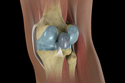
Knee Anatomy
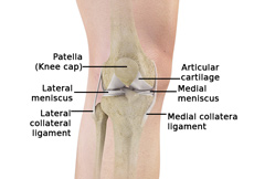
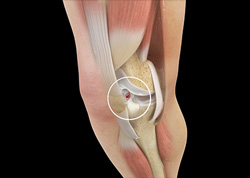
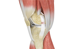
The knee is a complex joint made up of different structures including bones, tendons, ligaments and muscles. They all work together to maintain normal function and provide stability to the knee during movement.
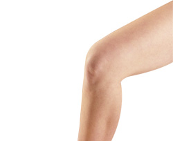
Having a well-functioning healthy knee is essential for our mobility and ability to participate in various activities. Understanding the anatomy of the knee enhances your ability to discuss and choose the right treatment procedure for knee problems with your doctor.
Physical examination of the knee
A complete physical examination of the knee is performed when you present to your doctor with a knee complaint. Both of your knees are examined and the results of the injured knee are compared to those of the healthy knee.
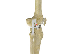
Know Your Knee
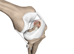
The knee joint is one of the most complex joints in the body. It consists of bones, ligaments, and muscles. The knee is made up of thefemur (thigh bone), the tibia (shin bone), and patella(kneecap). The meniscus, a soft cartilage between the femur and tibia, serves to cushion the knee and helps it absorb shock during motion.
-
Knee Pain
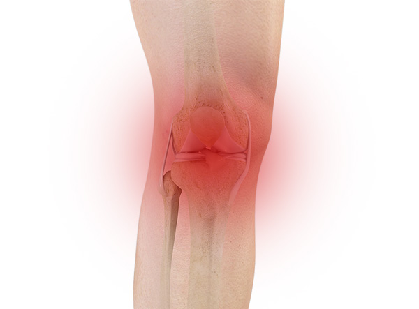
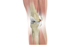
The knee is one of the largest joints in the body, formed by the lower end of the femur, upper end of the tibia and the patella or knee cap. Several ligaments and muscles attach to the bones of the knee joint to maintain normal motion of the joint.
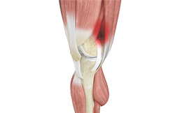
Anterior Knee Pain
Anterior knee pain is a characterized by a chronic pain over the front and center of the knee joint. It is common in athletes, active adolescents (especially girls) and overweight individuals. Anterior knee pain refers to a variety of conditions which include runner's knee or patellar tendinitis and chondromalacia of the patella.
Runner’s Knee
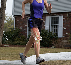
Runner's knee, also called patellofemoral pain syndrome refers to pain under and around your kneecap. Runner’s knee includes several medical conditions such as anterior knee pain syndrome, patellofemoral malalignment, and chondromalacia patella that cause pain around the front of the knee.
Osgood Schlatter Disease
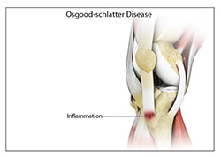
Osgood-Schlatter disease refers to a condition of an overuse injury that occurs in the knee region of growing children and adolescents. This is caused by inflammation of the tendon located below the knee cap (patellar tendon). Children and adolescents who participate in sports such as soccer, gymnastics, basketball and distance running are at higher risk of this disease.
Chondromalacia Patella
The patella, also called the kneecap is a small bone present on the front of your knee joint. The underside of the patella is covered by cartilage that allows smooth gliding of the knee with movement. Overuse or misalignment of the patella can cause wear and tear of the cartilage.
Jumper’s Knee
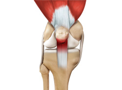
Jumper’s knee, also known as “patellar tendinitis" is an inflammation of the patellar tendon that connects your kneecap (patella) to your shinbone. This tendon helps in extension of the lower leg.
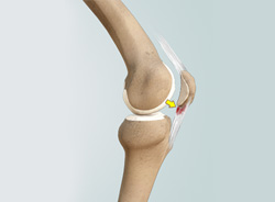
Bursitis
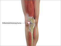
A bursa is a small fluid-filled sac found between soft tissues and bones. It lubricates and acts as a cushion to decrease friction between bones when they move. Bursitis refers to the inflammation and swelling of the bursa. Inflammation of the bursa in front of the kneecap (patella) is known as kneecap bursitis or prepatellar bursitis.
Baker’s Cyst
The knee consists of a fluid called synovial fluid, which reduces friction between the bones of the knee joint while you move your leg. Sometimes this fluid is produced in excess, resulting in its accumulation in the back of your knee. A Baker’s cyst or popliteal cyst is a fluid-filled swelling that develops into a lump behind the knee.
Iliotibial Band Syndrome
Iliotibial band syndrome is an overuse injury resulting from the inflammation of iliotibial band. Iliotibial band is a tough group of fibres that begins at the iliac crest of hip and runs along the outside of the thigh, to get attached to the outer side of the shin bone just below the knee joint. Its function is to coordinate with the thigh muscles and provide stability the knee joint.
Lateral Patellar Compression Syndrome
Lateral patellar compression syndrome refers to pain under and around your kneecap. It is a common complaint among runners, jumpers, and other athletes such as skiers, cyclists, and soccer players.
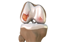
Osteochondritis Dissecans
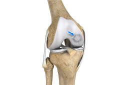
Osteochondritis dissecans is a joint condition in which a piece of cartilage, along with a thin layer of the bone separates from the end of the bone because of inadequate blood supply. The separated fragments are sometimes called “joint mice”. These fragments may be localised, or may detach and fall into the joint space causing pain and joint instability.
Shin Splints

“Shin splints” is used to describe the pain and inflammation of the tendons, muscles and bone tissue around the tibia or shine bone (a large bone in the lower leg). It occurs because of vigorous physical activity such as exercise or sports. The condition is also referred to as medial tibial stress syndrome (MTSS).
Knee Injuries and Tears
Pain, swelling and stiffness are the common symptoms of any damage or injury to the knee. If care is not taken during the initial phases of injury, it may lead to joint damage that may end up destroying your knee.
Knee Sprain
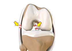
Knee sprain is a common injury that occurs from overstretching of the ligaments that support the knee joint. A knee sprain occurs when the knee ligaments are twisted or turned beyond its normal range causing the ligaments to tear.
ACL Tears
The anterior cruciate ligament, or ACL, is one of the major ligaments of the knee that is in the middle of the knee and runs from the femur (thigh bone) to the tibia (shin bone). It prevents the tibia from sliding out in front of the femur. Together with posterior cruciate ligament (PCL) it provides rotational stability to the knee.
MCL Tears
The medial collateral ligament (MCL) is the ligament that is located on the inner part of the knee joint. It runs from the femur (thighbone) to the top of the tibia (shinbone) and helps in stabilizing the knee. Medial collateral ligament (MCL) injury can result in a stretch, partial tear, or complete tear of the ligament.
MCL Sprain
The medial collateral ligament (MCL), a band of tissue present on the inside of your knee joint, connects your thigh bone and shin bone (bone of your lower leg). The MCL maintains the integrity of the knee joint and prevents it from bending inward.
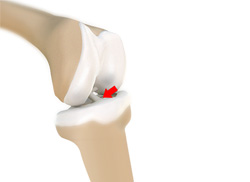
Meniscal Injuries
The knee is one of the most complex and largest joint in the body, and is more susceptible to injury. Meniscal tears are one among the common injuries to the knee joint. It can occur at any age, but are more common in athletes playing contact sports.
Meniscal Tears
Meniscus tear is the commonest knee injury in athletes, especially those involved in contact sports. A suddenly bend or twist in your knee cause the meniscus to tear. This is a traumatic meniscus tear. Elderly people are more prone to degenerative meniscal tears as the cartilage wears out and weakens with age.
Ligament Injuries
The knee is a complex joint which consists of bone, cartilage, ligaments and tendons that make joint movements easy and at the same time more susceptible to various kinds of injuries.
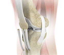
Multiligament Instability

The knee is a complex joint of the body which is vital for movement. The four major ligaments of the knee are anterior cruciate ligament, posterior cruciate ligament, medial collateral ligament and lateral collateral ligament. They play an important role in maintaining the stability of the knee.
Multi-ligament Injuries
Ligaments are the fibrous tissue bands connecting the bones in the joint and stabilizing the joint. The knee has four major ligaments – the anterior cruciate ligament, posterior cruciate ligament, lateral collateral ligament, and medial collateral ligament. There is also a "fifth ligament" of the knee which is the posterior lateral corner, which is a complex of multiple small ligaments and muscles.
Knee Arthritis
Arthritis is a general term covering numerous conditions where the joint surface or cartilage wears out. The joint surface is covered by a smooth articular surface that allows pain free movement in the joint. This surface can wear out for several reasons; often the definite cause is not known.
Osteoarthritis
Osteoarthritis, also called degenerative joint disease is the most common form of arthritis. It occurs most often in older people. This disease affects the tissue covering the ends of bones in a joint (cartilage). In a person with osteoarthritis, the cartilage becomes damaged and worn out causing pain, swelling, stiffness and restricted movement in the affected joint.
Patellar Dislocation/Patellofemoral Dislocation
Patella (knee cap) is a protective bone attached to the quadriceps muscles of the thigh by quadriceps tendon. Patella attaches with the femur bone and forms a patellofemoral joint. Patella is protected by a ligament which secures the kneecap from gliding out and is called as medial patellofemoral ligament (MPFL).
Patellar tendinitis
Patellar tendinitis, also known as “jumper’s knee” is an inflammation of the patellar tendon that connects your kneecap (patella) to your shinbone. This tendon helps in extension of the lower leg. Patellar tendinitis usually results from repetitive trauma or overuse, particularly from sports activities involving jumping such as basketball or volleyball.
PCL Injuries
Posterior cruciate ligament (PCL), one of four major ligaments of the knee are situated at the back of the knee. It connects the thighbone (femur) to the shinbone (tibia). The PCL limits the backward motion of the shinbone.
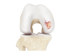
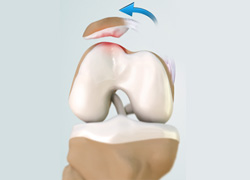
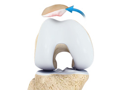
Chondral
Articular or hyaline cartilage is the tissue lining the surface of the two bones in the knee joint. Cartilage helps the bones move smoothly against each other and can withstand the weight of the body during activities such as running and jumping. Articular cartilage does not have a direct blood supply to it so has less capacity to repair itself.
Patellar Instability
Patellar (knee cap) instability results from one or more dislocations or partial dislocations (subluxations). Patella is the small piece of bone in front of the knee that slides up and down the femoral groove (groove in the femur bone) during bending and stretching movements.
Patella Femoral Dislocation
Patella (knee cap) is a protective bone attached to the quadriceps muscles of the thigh by quadriceps tendon. Patella attaches with the femur bone and forms a patellofemoral joint. Patella is protected by a ligament which secures the kneecap from gliding out and is called as medial patellofemoral ligament (MPFL).
Patellofemoral Instability
The knee can be divided into three compartments: patellofemoral, medial and lateral compartment. The patellofemoral compartment is the compartment in the front of the knee between the knee cap and thigh bone. The medial compartment is the area on the inside portion of the knee, and the lateral compartment is the area on the outside portion of the knee joint.
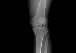
Patella Fracture
The knee cap or patella is the largest sesamoid bone in the body and one of the components of the knee joint, present at the front of the knee. The under surface of the kneecap and the lower end of the femur are coated with articular cartilage, which helps in smooth movement of the knee joint. The knee cap protects the knee and provides attachment to various muscle groups of the thigh and leg.
Quadriceps Tendon Rupture
Quadriceps tendon is a thick tissue located at the top of the kneecap. The quadriceps tendon works together with the quadriceps muscles to allow us to straighten our leg. The quadriceps muscles are the muscles located in front of the thigh.
Patella Tendon Rupture
Patella tendon rupture is the rupture of the tendon that connects the patella (knee cap) to the top portion of the tibia (shin bone). The patellar tendon works together with the quadriceps muscle and the quadriceps tendon to allow your knee to straighten out.
Lateral Meniscus Syndrome
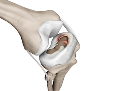
The knee joint is formed by the union of two bones, namely the femur (thigh bone) and the tibia (lower leg bone). At the junction of these two bones is a cartilage called the meniscus, which acts as a shock absorber. There are two menisci – the lateral and medial menisci.
Medial Meniscus Syndrome
Of the menisci within the knee, it is the medial that is more easily injured. Differences in the anatomical attachments of the medial meniscus compared to the lateral mean that the medial meniscus becomes distorted during combined flexion and rotation movements in a manner not experienced on the lateral side.
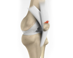
Tibial Eminence Spine Avulsions
Tibial eminence spine avulsion fracture is avulsion (tear away) of the tibial eminence (an extension on the bone for attachment of muscles) which most commonly involves the anterior cruciate ligament (ACL) insertion site.
Osteonecrosis of the Knee
Osteonecrosis is a condition in which death of a section of bone occurs because of lack of blood supply to it. It is one of the most common causes of knee pain in older women. Women over the age of 60 years of age are commonly affected, three times more often than men.
Knee Angular Deformities
Angular deformities of the knee are common during childhood and usually are variations in the normal growth pattern. Angular deformity of the knee is a part of normal growth and development during early childhood.