Dr Julian Yu is a specialist orthopaedic surgeon performing distal realignment procedures for conditions affecting the patella in the knee.

What is a tibial tubercle transfer?
Tibial tubercle transfer, also known as a distal alignment procedure, is used to realign the patella (kneecap) and treat patellar instability, chronic dislocation, or maltracking of the kneecap. This procedure helps reduce pain and improve the function of the knee, particularly in patients with conditions like patellar instability, recurrent dislocation, or patellofemoral pain syndrome.
When is a tibial tubercle transfer considered?
TTT may be considered when conservative treatments such as physiotherapy, bracing, and medications fail. Common conditions that may benefit from TTT include:
- Chronic patellar dislocation or subluxation
- Patellofemoral arthritis or cartilage damage
- Pain due to patellar malalignment or maltracking
- Congenital alignment issues
- Increased tibial tubercle–trochlear groove (TT-TG) distance
What are the types of distal realignment procedures?
- Maquet procedure
In this procedure, the tibia tubercle is cut, keeping the patellar tendon attachment intact. The tubercle is elevated by wedging the loosened piece of bone using a bone block. This procedure cannot move the tendon and tubercle medially (towards the inner aspect of the knee). - Elmslie-Trillat procedure
This is a procedure like Maquet procedure, but the tendon and tubercle can be moved medially. - Fulkerson procedure
In this procedure, the tibia tubercle is moved more towards the inner aspect of the knee. This is achieved by breaking the bone into sharp pieces (splintered) which allows the bit of bone and the tendon to move more medially. After the procedure bits of bone are held in place using screws. - Hauser procedure
In this procedure, the tibia tubercle is moved medially, but not moved forward (anterior). Because of the shape of the tibia, the tubercle may shift its position more posteriorly and the patella may press down causing pain. - Roux-Goldthwait procedure
It is a distal realignment procedure where the patellar tendon is split vertically. The lateral half of the patellar tendon is pulled under the inner half (medial) and attached to tibia. This pulls the patella over to the centre and helps prevent excess lateral shift.
After a distal realignment procedure
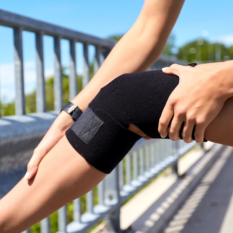
Most people stay in the hospital for 1 to 2 days after surgery. During this time, Dr Yu will focus on managing pain and checking your early recovery. You will wear a knee brace to keep your leg straight and protect the area where the surgery was done. Crutches will help you move around without putting weight on your healing leg, and you may need them for several weeks.
Physiotherapy usually starts soon after the surgery. It helps you slowly get your movement back, build up your strength, and improve how your knee works.
It can take about 4 to 6 months before you can go back to sports or other high-impact activities, depending on how well your recovery goes.
For all appointments and enquiries, please phone (02) 8045 5688
Monday to Friday 9am–5pm
Frenchs Forest
Peninsula Orthopaedics
Suite 20, Level 7
Northern Beaches Hospital
105 Frenchs Forest Rd
Frenchs Forest NSW 2086
Chatswood
Orthopaedic & Arthritis Specialist Centre
Level 2, Gallery Arcade
445 Victoria Avenue
Chatswood NSW 2067
© 2018– Dr Julian Yu | Privacy Policy | Disclaimer | Website by:

