Dr Julian Yu is a specialist orthopaedic surgeon performing tibial tubercle osteotomy for conditions affecting the patella in the knee.

What is a tibial tubercle osteotomy?
Tibial tubercle osteotomy is used to realign the patella (kneecap) and improve its tracking within the groove at the end of the femur (trochlea). This procedure is often recommended for individuals experiencing chronic kneecap instability, recurrent patellar dislocations, or anterior knee pain due to abnormal patellar tracking.
When is a tibial tubercle osteotomy considered?
TTO may be considered when conservative treatments such as physiotherapy, bracing, and medications fail. Common conditions that may benefit from TTO include:
- Recurrent patellar dislocations or instability
- Patellofemoral pain syndrome
- Patella alta (high-riding kneecap)
- Chondromalacia patella (softening of cartilage under the kneecap)
- Maltracking of the patella
- Cartilage damage in the front of the knee
What happens during a tibial tubercle ostetomy?
Tibial tubercle osteotomy and transfer is performed through an incision at the front of the leg, just below the kneecap. During the procedure, a 8–10 cm long incision is made about 1 cm inside (medial) to the tibial tubercle. Using an oscillating saw, a cut is made on the inner side of the tubercle, followed by a tapered distal cut, which helps reduce the risk of fracturing the tibia. A cut is also made on the upper (proximal) side using surgical tools like a curved osteotome or reciprocating saw. Dr Yu carefully cuts through the bone's outer layer (cortex) while keeping the outer side's soft tissue (lateral periosteum) intact. This preserved tissue acts as a hinge to keep the cut bone segment attached.
The resulting bone segment typically measures over 2 cm wide, more than 1 cm thick, and 8–10 cm long. It includes the entire attachment site of the patellar tendon. The cut bone is gently lifted to allow access to the inner part of the tibia (medullary canal).
Under direct visual guidance—and sometimes with arthroscopy to check kneecap movement—the surgeon repositions the bone to improve patellar alignment. Once the best position is found, the bone segment is secured in place with screws. These screws can be removed later if they cause discomfort.
After a tibial tubercle ostetomy
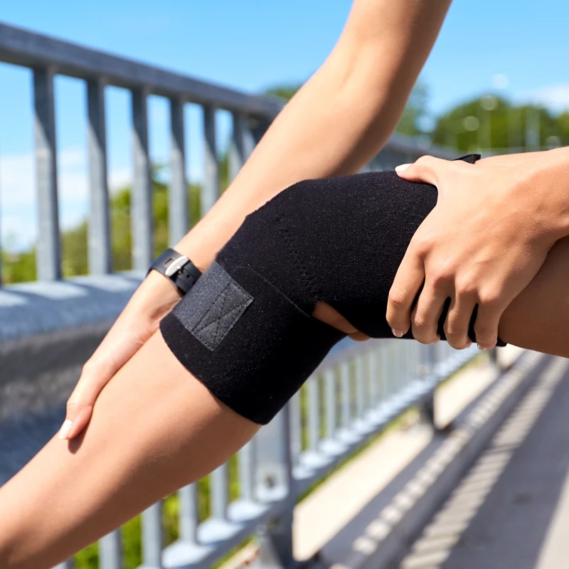
You may experience mild to moderate knee pain for a few days or weeks following the surgery. To help manage this, Dr Yu will prescribe oral pain medication. Keeping your leg elevated and applying an ice pack for 20 minutes at a time can help reduce both swelling and discomfort. You’ll be given a leg brace, which should only be taken off while sitting with your leg elevated or when using a continuous passive motion (CPM) machine.
For all appointments and enquiries, please phone (02) 8045 5688
Monday to Friday 9am–5pm
Frenchs Forest
Peninsula Orthopaedics
Suite 20, Level 7
Northern Beaches Hospital
105 Frenchs Forest Rd
Frenchs Forest NSW 2086
Chatswood
Orthopaedic & Arthritis Specialist Centre
Level 2, Gallery Arcade
445 Victoria Avenue
Chatswood NSW 2067
© 2018– Dr Julian Yu | Privacy Policy | Disclaimer | Website by:

